What Is a Retinal Vein Occlusion?
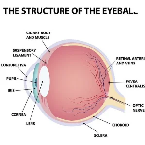
The retina is the light sensing tissue that lines the inside back part of our eye. A retinal vein occlusion means that a vein in the retina has become blocked. The veins drain blood out of the eye and return it to the heart.
Blockage or occlusion in the vein prevents adequate blood flow in the affected area. The walls of the vein leak blood and excess fluid into the retina, usually resulting in blurred vision if the leakage involves the central portion of the retina known as the macula.
What Are the Types of Retinal Vein Occlusion?
There are three types of retinal vein occlusion:
Hemiretinal Vein Occlusion (HRVO):
A main branch from the central retinal vein has become blocked, leading to engorged vessels, leakage and hemorrhage in one-half of the retina.
Central Retinal Vein Occlusion (CRVO):
The main retinal vein has become blocked in this condition, leading to engorged vessels, leakage, and hemorrhage throughout the retina.
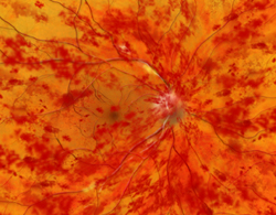
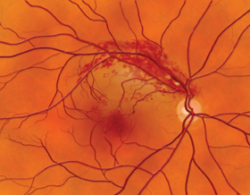
Branch Retinal Vein Occlusion (BRVO):
A smaller branch retinal vein has become blocked, leading to engorged vessels, leakage, and hemorrhage in a wedge-shaped portion of the retina.

Who Is At Risk for a Retinal Vein Occlusion?
Retinal vein occlusions are more common in people who have:
- Glaucoma (abnormal eye pressure)
- Diabetes
- Age-related vascular (blood vessel) disease
- High blood pressure
- Blood disorders

What Are the Symptoms of Retinal Vein Occlusion?
- Blurred vision is the main symptom of retinal vein occlusion. It occurs when the excess fluid leaking from the vein collects in the macula. The macula is the central area of the retina which is responsible for our central, detailed vision. If the macula swells with excess fluid (macular edema), vision blurs.
- Floaters can appear as spots which interfere with vision. When retinal blood vessels are not working properly, the retina may grow abnormal blood vessels (neovascularization), which are fragile. They can bleed or leak fluid into the vitreous—the gel-like fluid that fills the center of the eye—causing the floaters.
- Pain in the eye sometimes occurs as a complication of severe CRVO. It is caused by excessive eye pressure called neovascular glaucoma.
What Tests Might Be Ordered?
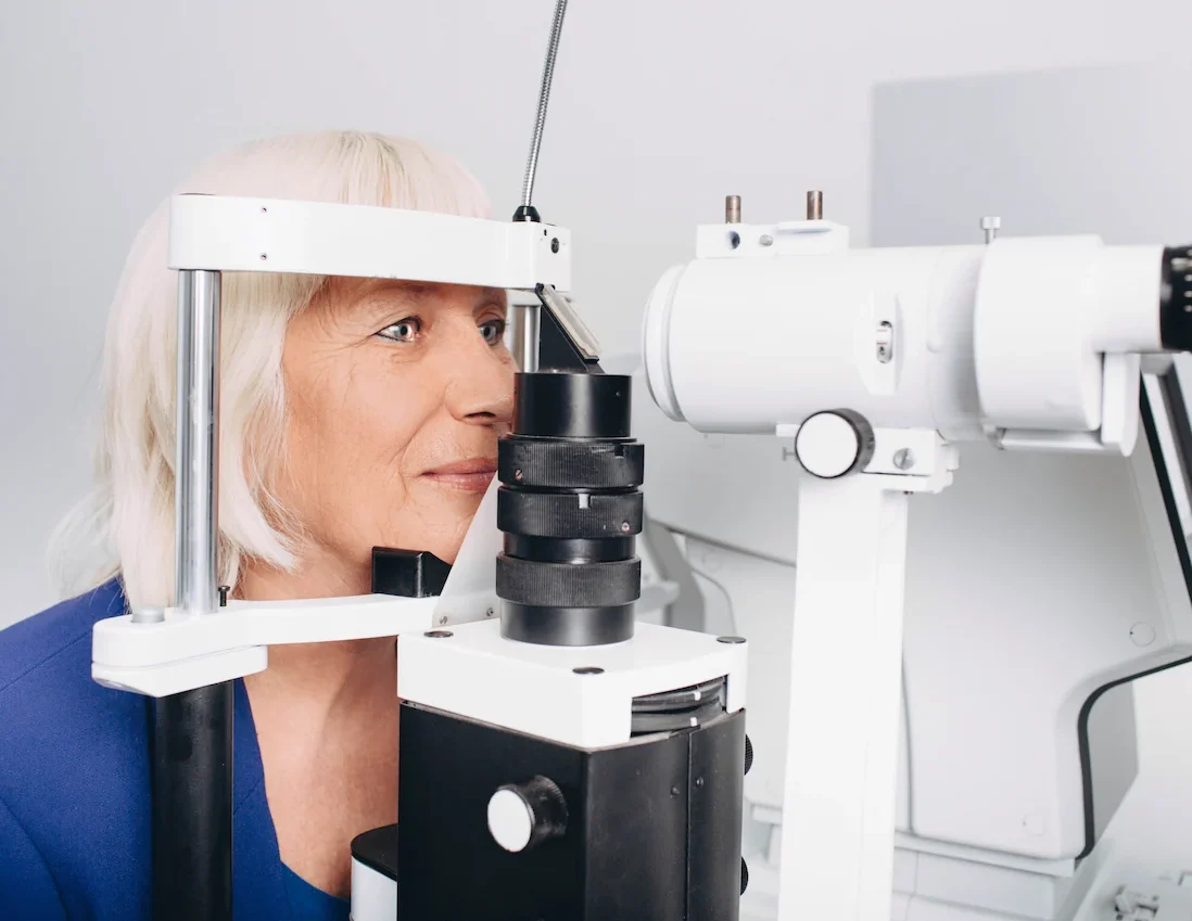
After a complete eye examination, your doctor may order blood tests and/or a test of the retinal circulation called fluorescein angiography (FA). To perform an FA, a vegetable-based dye (fluorescein) is injected into your arm, and then special photos are taken of the inside of your eye. As the dye passes to your eye, more photos are taken to document blood flow and potential leakage. Other specialized testing such as Optical Coherence Tomography (OCT) is sometimes useful to quantify swelling, gauge treatment and aid in visual prognosis. Your doctor may also suggest a visit to your family physician to discover and manage any associated medical problems.
What Treatment Is Available?
Presently, there is no cure for retinal vein occlusion. Your doctor may recommend a period of observation, since hemorrhages and excess fluid may subside on their own.
- Laser Surgery improves sight in some patients with BRVO and macular edema, but vision does not usually return to normal. A large, randomized, controlled clinical trial demonstrated that patients with CRVO and macular edema are not helped by laser surgery. Laser surgery is very effective in preventing vitreous hemorrhage and neovascular glaucoma. However, it does not remove hemorrhage or cure neovascular glaucoma once they are already present. It is best to treat people at risk for these complications before they occur. Your doctor will decide whether laser surgery is appropriate for you. Frequent follow-up examinations are essential. You should make sure that any associated medical condition is treated by your regular physician.
- Intraocular Injections are being used to treat patients with BRVO, HRVO and CRVO. Although just now being studied in randomized, controlled clinical trials, the doctors at Retina Specialty Institute offer injections of intraocular steroids and/or anti-blood vessel medications to patients who have poor blood flow or are unresponsive to grid laser treatment. Unfortunately, multiple injections are sometimes necessary to control macular edema. A recent advance in a slow-release steroid implant shows promise in these patients.
Why Are Regular Medical Eye Examinations Important for Everyone?
Eye disease can occur at any age. Many eye diseases do not cause symptoms until the disease has done damage. Since most blindness is preventable if diagnosed and treated early, regular medical examinations by an eye doctor are very important.
Retina Specialty Institute has been involved in multiple clinical trials, including studies involving retinal vein occlusions. Your retinal surgeon or our staff can answer any questions you may have concerning retinal vein occlusions.

Schedule a Consultation
At Retina Specialty Institute, our number one priority has always been and will always be offering exceptional eye care to our patients in a safe and professional environment. Contact us today for an appointment. We have a wide range of amazing, state-of-the-art locations.
The doctors at Retina Specialty Institute have either authored or reviewed and approved this content.
Page Updated:

